
Complete Guide to Ankle Joint Damage – Diagnosis, Treatment and Recovery
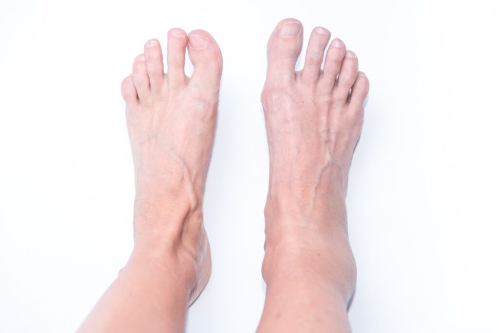
What is the Ankle Mortise?
The ankle mortise refers to the socket formed by the tibia and fibula that securely houses the talus bone. When this structure is disrupted, the joint can lose its alignment and stability.
Disruption of the ankle mortise and talar dome injuries are serious conditions that compromise the ankle joint’s stability and function.
The ankle mortise is formed by the tibia and fibula, which together create a secure socket for the talus bone. This structure allows the ankle to function as the tibiotalar joint, providing smooth up-and-down movement of the foot.
Key functions include:
- Holding the talus in correct alignment
- Providing stability for walking and running
- Distributing weight evenly through the ankle joint
When intact, the mortise is essential for everyday mobility and balance.
Talar dome injuries, on the other hand, involve damage to the cartilage surface of the talus within the joint. These injuries cause pain, swelling, and mechanical symptoms. Both conditions can significantly impact walking, sporting activities, and long-term joint health if they are not identified and managed appropriately.
What is Disruption of the Ankle Mortise?
Disruption occurs when the talus no longer sits properly within the mortise, often due to injury. This may result from fractures, syndesmotic ligament damage, or significant trauma.
- The joint becomes unstable and painful.
- Weight-bearing is often difficult or impossible.
- If not treated, the disruption increases the risk of early arthritis.
What are Talar Dome Injuries?
The talar dome is the smooth, cartilage-covered surface on the top of the talus. Injuries here are commonly referred to as osteochondral lesions, affecting bone and cartilage.
These injuries may be caused by:
- Severe ankle sprains
- Repeated ankle instability
- Direct trauma or impact to the joint
Typical symptoms include deep ankle pain, swelling and stiffness. Sometimes a catching or locking sensation occurs when moving the ankle.
What are the Types of Ankle Mortise Disruption?
Ankle mortise disruption can take several forms:
- Syndesmotic injuries – damage to the ligaments between the tibia and fibula, also known as high ankle sprains.
- Malleolar fractures – breaks of the medial or lateral malleolus that compromise joint stability.
- Joint incongruity – where the talus no longer aligns correctly within the mortise, altering ankle mechanics.
These injuries are considered serious and usually require prompt assessment and treatment.
What are the Types of Talar Dome Injuries?
There are several ways the talar dome can be damaged:
- Osteochondral defects (OCDs) – small areas of cartilage and underlying bone damage.
- Talar dome fractures – breaks in the talus bone surface.
- Cartilage lesions – tears or degeneration of the smooth cartilage lining the dome.
Each of these injuries interferes with ankle function and can progress to arthritis if untreated.
How Common are Ankle Mortise and Talar Dome Injuries?
Disruption of the ankle mortise is most often seen in young, active individuals following high-energy trauma such as sporting injuries or falls. Talar dome injuries are less common, but they can be underdiagnosed because their symptoms resemble those of a severe ankle sprain.
They are most commonly seen in:
- Athletes involved in running, jumping, or contact sports
- Adults who have suffered ankle fractures or repeated sprains
Although not the most frequent ankle injuries, both conditions are clinically significant due to their potential to cause long-term pain, instability, and early arthritis.
Symptoms and Causes
What Causes Ankle Mortise Disruption?
Ankle mortise disruption usually occurs after significant trauma. The most common mechanism is a high-energy injury, where the bones and ligaments holding the talus in place are damaged.
Typical causes include:
- High-impact sports accidents or collisions
- Falls from a height
- Motor vehicle accidents
- Complex ankle fracture patterns, particularly those involving the tibia and fibula
These injuries often require urgent assessment due to the risk of long-term joint damage.
What Causes Talar Dome Injuries?
Talar dome injuries are commonly linked to twisting injuries of the ankle, particularly severe sprains. Repetitive trauma can gradually weaken the cartilage of the talus, while in some cases, the blood supply to the bone is impaired, leading to osteonecrosis.
Key causes include:
- Severe ankle sprains or repeated sprains
- Chronic ankle instability
- Repetitive impact from sports such as basketball, football, or running
- Degenerative changes or reduced blood flow to the talus (osteonecrosis)
What are Ankle Mortise Disruption Risk Factors?
Certain individuals are more likely to sustain an ankle mortise injury. The risk increases with:
- Participation in high-impact activities or contact sports
- A history of previous ankle injuries
- Anatomical variations, such as weak ligament support or bone alignment differences
What are Talar Dome Injury Risk Factors?
Talar dome injuries are more likely to occur in athletes and people who place repetitive stress on the ankle. Risk factors include:
- Regular participation in running, jumping, or pivoting sports
- Chronic ankle instability following untreated sprains
- Repetitive loading of the ankle joint without adequate recovery
What are the Signs and Symptoms of Ankle Mortise Disruption?
Ankle mortise disruption presents dramatically and is often obvious at the time of injury. Common symptoms include:
- Severe pain in the ankle joint
- Visible deformity or abnormal alignment
- Marked instability of the ankle
- Inability to bear weight or walk
These injuries usually require urgent medical assessment.
What are the Signs and Symptoms of Talar Dome Injuries?
Talar dome injuries may not be immediately apparent, and symptoms often mimic those of a severe sprain. Patients typically report:
- Deep ankle pain that worsens with activity
- Swelling and stiffness in the joint
- Catching, locking, or clicking sensations within the ankle
- Pain that flares during sport or weight-bearing activities
What are the Complications of Ankle Mortise Disruption and Talar Dome Injuries?
If not treated properly, disruption of the ankle mortise and talar injuries can lead to serious long-term consequences. These include:
- Post-traumatic arthritis caused by abnormal joint loading
- Chronic ankle instability and weakness
- Malunion of fractures, where bones heal in poor alignment
Diagnosis and Tests
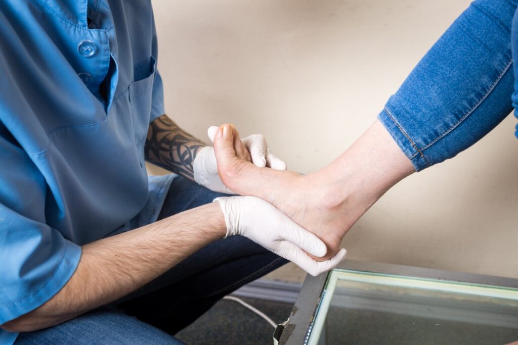
How are Ankle Mortise Disruptions Diagnosed?
Ankle mortise disruptions are usually identified through a detailed history and clinical examination. A provider will assess the ankle for pain, swelling, and deformity and check whether the joint feels stable under stress. They may also evaluate the ability to bear weight and look for signs of ligament damage or associated fractures.
How are Talar Dome Injuries Diagnosed?
Talar dome injuries can be more challenging to diagnose. A clinical examination often reveals deep ankle pain, swelling, and tenderness, which overlap with common ankle sprains. Special clinical tests may be used to check for instability or joint locking, though imaging is usually required to confirm the diagnosis.
Which Tests do Providers Use to Diagnose Ankle Mortise Disruption?
Imaging plays a crucial role. Standard ankle X-rays help detect fractures and alignment issues within the mortise. In more complex cases, CT scans provide detailed views of fracture patterns. Stress X-rays may be recommended to assess joint stability and identify subtle widening that indicates ligament injury.
Which Tests do Providers Use to Diagnose Talar Dome Injuries?
Because talar dome injuries often involve cartilage or bone bruising that may not show up on plain X-rays, advanced imaging is required. MRI scans are the gold standard as they provide detailed images of cartilage and bone integrity. CT arthrography may be used to outline cartilage defects, and in some cases, arthroscopy (a minimally invasive surgical camera inspection) is performed to directly visualise the joint surface.
Ankle Mortise X-ray Assessment
X-rays are the first-line tool for evaluating ankle mortise injuries. Providers look for abnormal joint spacing, shifts in alignment, or signs of a fracture. Specific measurements, such as the medial clear space and tibiofibular overlap, help doctors determine whether the mortise remains intact or has widened due to ligament disruption.
Talar Dome MRI Imaging
MRI is the preferred imaging test for talar dome lesions. It provides detailed views of the cartilage, subchondral bone, and any associated oedema or cystic changes. This information is essential for grading the severity of the injury and planning treatment, whether conservative or surgical.

Specific Considerations
Acute vs Chronic Ankle Mortise Disruption
An acute ankle mortise disruption occurs immediately after a significant traumatic injury, such as a fall, sports tackle, or motor vehicle accident. These injuries typically require urgent medical attention to restore joint alignment and prevent long-term complications. Chronic disruptions may develop if the initial injury is missed or inadequately treated. In chronic cases, patients often present with persistent pain, instability, and arthritis, which can complicate treatment and limit outcomes.
Access Ortho treats acute musculoskeletal injuries in the urgent care fracture clinics.
Acute vs Chronic Talar Dome Injuries
Talar dome injuries may present acutely after a significant ankle sprain or fracture, with patients experiencing sharp pain and swelling. However, in many cases, symptoms evolve gradually. Chronic lesions may present months after the initial trauma, with ongoing pain, catching or locking sensations, and reduced performance. These delayed presentations make diagnosis more challenging, often requiring advanced imaging to identify cartilage or bone damage that has failed to heal.
Access Ortho offers rapid appointments for acute injuries, with fracture clinics in Brisbane and Ipswich.
Ankle Mortise Disruption in High-Energy Trauma
High-energy accidents, such as car crashes or falls from height, can cause severe ankle mortise disruption. These injuries are frequently associated with other fractures or polytrauma, requiring a coordinated ambulance and hospital emergency response.
Initial management focuses on stabilising the patient, controlling pain, and restoring blood flow to the limb. Surgical repair is often needed once the patient is stable.
Talar Dome Injuries in Athletes
For athletes, talar dome injuries can significantly impact performance. Even small cartilage defects can cause ongoing pain during running, jumping, or pivoting movements. A rapid, accurate diagnosis is crucial, as untreated lesions can limit training and competition. Treatment is often tailored to preserve joint function and minimise downtime, with rehabilitation playing a central role in return-to-play planning.
Paediatric Ankle Mortise and Talar Dome Injuries
Children and adolescents require special consideration because of open growth plates. Ankle mortise injuries in this group may involve the physis (growth plate), which can affect long-term bone development if not managed appropriately. Talar dome injuries may also present differently in children, with subtle symptoms but the potential for long-lasting joint problems. Careful imaging and paediatric-specific treatment approaches are essential to protect growth and restore function.
Access Ortho treats adults and children in their private fracture clinics.
Associated Injuries with Ankle Mortise Disruption
Ankle mortise injuries often occur alongside damage to surrounding structures. These may include ligament tears (such as the syndesmotic ligaments), nerve stretching or compression injuries, and, in severe cases this could lead to compartment syndrome. Compartment syndrome occurs where swelling restricts blood supply to the lower leg. Identifying and treating these associated injuries early is critical to prevent long-term disability.
Management and Treatment
How are Ankle Mortise Disruptions Treated?
Treatment depends on how severe the disruption is and the stability of the ankle joint. Stable injuries may be managed with immobilisation, while unstable or displaced injuries typically require surgery to restore alignment and prevent long-term complications such as arthritis. Surgical decision-making will depend on the fracture pattern, degree of displacement, and syndesmotic integrity.
If surgery is required, the Access Ortho team will assist in arranging it.
How are Talar Dome Injuries Treated?
Talar dome injuries can be managed either conservatively or surgically, depending on lesion size, depth, and symptoms. Small or stable injuries may improve with activity modification, bracing, and physiotherapy. Larger or unstable lesions, or those that fail conservative care, often require surgery to restore cartilage and prevent chronic pain or joint degeneration.
If surgery is required, the orthopaedic team at Access Ortho can help arrange this.
Non-Surgical Treatment for Ankle Mortise Disruption
Conservative management may be suitable for non-displaced or minimally unstable injuries. Treatment may include:
- Immobilisation in a cast or boot to protect the joint
- Restricted weight-bearing until stability is confirmed
- Close follow-up imaging to monitor alignment and healing
- Physiotherapy is introduced to gradually restore movement, strength, and stability once healing has progressed
Non-Surgical Treatment for Talar Dome Injuries
Stable or less severe talar dome lesions may improve without surgery. Non-surgical options include:
- Rest and modifications to activity to reduce stress on the joint
- Supportive bracing to limit excessive movement and protect the ankle
- Physiotherapy focusing on strength, mobility, and proprioception
- Injection therapy (e.g., platelet-rich plasma or corticosteroid injections) in selected cases to reduce inflammation and promote healing
Surgical Treatment for Ankle Mortise Disruption
When the ankle mortise is unstable or displaced, surgery is required. The standard approach is open reduction and internal fixation (ORIF), which uses plates and screws to restore proper joint alignment. In more complex cases, reconstruction of the syndesmosis (ligament complex between tibia and fibula) may also be necessary to restore stability.
Surgical Treatment for Talar Dome Injuries
Surgical approaches depend on the size and nature of the lesion. Options include arthroscopic debridement (removing loose fragments), microfracture (stimulating new cartilage growth by creating small holes in the bone), or advanced cartilage restoration procedures. These minimally invasive surgeries aim to relieve pain and preserve joint function.
Ankle Mortise Reconstruction Surgery
In severe or chronic cases, complex reconstruction may be required. This may involve the use of plates, screws, suture-button devices, or even external fixation to stabilise the ankle. Reconstruction addresses both the bony alignment and ligament integrity to restore joint stability and reduce the risk of long-term arthritis.
Talar Dome Cartilage Restoration Procedures
Cartilage restoration techniques are used for large or persistent talar dome lesions. Options include osteochondral grafting (transplanting cartilage and bone plugs), autologous chondrocyte implantation (ACI) (culturing a patient’s own cartilage cells for reimplantation), and newer biologic or scaffold-based procedures. These techniques aim to restore joint surface integrity and delay degenerative change.
Rehabilitation After Ankle Mortise Surgery
Rehabilitation is critical for recovery. Early phases focus on protecting the repair with limited or non-weight-bearing and controlled motion exercises. Gradual weight-bearing is introduced under medical guidance, followed by progressive strengthening, balance training, and return-to-activity protocols. Recovery timelines vary but often extend over several months.
Rehabilitation After Talar Dome Surgery
Post-operative rehabilitation for talar dome injuries emphasises gradual activity progression. Early stages involve protected weight-bearing and gentle range-of-motion work. Later phases incorporate strengthening, proprioception training, and sport-specific drills. Return to full athletic activity may take 6–12 months, depending on the size of the lesion and the surgical technique used.
Prevention
How can I Prevent Ankle Mortise Disruption?
While not all ankle injuries can be avoided, certain strategies reduce the risk:
- Wear supportive footwear during sports or high-risk activities
- Use ankle braces or taping if you have a history of instability
- Strengthen the lower limb muscles especially around the ankle and lower leg for joint protection
- Avoid uneven surfaces where possible to reduce the chance of twisting injuries
How can I Prevent Talar Dome Injuries?
Talar dome injuries often result from repetitive ankle trauma. Preventative measures focus on stability and joint protection, including:
- Regular ankle stability training (balance, proprioception, and agility drills)
- Addressing ankle sprains early to prevent chronic instability
- Maintaining flexibility in the calf and foot muscles to reduce joint loading
Ankle Injury Prevention in Sports
Athletes face higher risks of ankle injuries. Protective measures include:
- Structured warm-ups and mobility drills before training or competition
- Progressive strength and conditioning programs tailored to sport demands
- Appropriate footwear for the playing surface
- Use of ankle taping or braces for those with previous injuries
Proper Ankle Conditioning and Strengthening
A strong and well-conditioned ankle is less prone to injury. Helpful strategies include:
- Calf raises, theraband exercises, and balance training to build stability
- Agility ladder drills to improve movement control
- Core and hip strengthening, which reduces stress on the ankle joint
- Consistent rehabilitation following sprains to avoid long-term weakness
Long-term Outcomes of Ankle Joint Injuries
- Some patients develop post-traumatic arthritis due to joint surface damage
- Functional limitations such as stiffness, pain, or instability may persist in severe cases
- Most patients achieve a good quality of life with early, appropriate treatment and adherence to rehabilitation
Outlook / Prognosis
What can I expect with an Ankle Mortise Disruption?
Recovery depends on the degree of the injury and whether surgery is required.
- With appropriate treatment, most patients regain good ankle stability.
- Severe disruptions may increase the risk of long-term complications such as arthritis.
What can I expect with Talar Dome Injuries?
Outcomes vary depending on the size and stability of the lesion.
- Small or stable injuries may heal successfully with conservative treatment.
- Larger or chronic lesions often require surgery, and recovery can be more complex.
- Athletes may face more extended rehabilitation, but often return to sport following structured treatment and physiotherapy.
What is the Recovery Time from Ankle Mortise Surgery?
Recovery occurs in phases:
- Initial healing: 6–8 weeks of immobilisation and limited weight-bearing
- Rehabilitation: several months of physiotherapy to restore motion, strength, and balance
- Return to activity: 4–6 months for daily activities, with full recovery sometimes taking 9–12 months
What is the Recovery Time from Talar Dome Surgery?
Rehabilitation timelines differ based on the surgical approach and lesion severity.
- Protected weight-bearing: typically 6–8 weeks post-surgery
- Physiotherapy progression: strength and proprioception work begins around 2–3 months
- Return to sport: usually within 6–12 months, depending on healing response and surgical technique used
When should I see an Orthopaedic Specialist for Ankle Problems?
You should seek a specialist opinion if you experience:
- Persistent or severe ankle pain after an injury
- Inability to bear weight or obvious deformity
- Ongoing swelling, instability, or locking of the ankle
- Lack of improvement after a period of rest and conservative care
Access Ortho has urgent care fracture clinics in Brisbane and Ipswich, which offer rapid appointments for acute injuries including fractures, sprains, and strains.
When to Seek Immediate Medical Attention
Ankle Injury Emergency Signs
Some ankle injuries require urgent medical assessment. Seek emergency care if you experience:
- Severe pain or swelling that does not settle with rest
- Inability to bear weight or walk even a few steps
- Visible deformity or abnormal joint alignment
- Numbness, tingling, or loss of circulation in the foot
- Rapidly increasing swelling or tightness in the leg (possible compartment syndrome)
Fracture clinics play an essential role in assessing and treating these injuries promptly. They provide expert diagnosis, imaging, and specialist-led treatment to reduce complications and optimise recovery.
Suspected Ankle Mortise Disruption First Aid
If you suspect an ankle mortise disruption:
- Stop activity immediately and avoid putting weight on the injured ankle.
- Immobilise the joint using a splint or supportive device if available.
- Apply ice and keep the ankle elevated to reduce swelling.
- Seek urgent assessment at a fracture clinic or emergency department.
At Access Ortho, patients can bypass long hospital waits and receive rapid care from orthopaedic specialists. The clinic provides rapid appointments, imaging referrals, and early treatment planning for ankle mortise disruptions and other complex injuries.
What is a Fracture Clinic?
A fracture clinic is a specialised medical facility where patients with bone, joint, and cartilage injuries are diagnosed, treated, and monitored. These clinics bring together:
- Orthopaedic surgeons with expertise in complex fractures and joint injuries
- Nurse practitioners trained in acute musculoskeletal care and ongoing management
- Rapid imaging referrals and follow-up care to streamline diagnosis and treatment
Access Ortho is a private urgent fracture clinic offering rapid assessment and treatment for ankle injuries, including ankle mortise disruptions and talar dome lesions. With shorter waiting times and direct access to orthopaedic expertise, patients can receive the right care quickly.
When should I go to a Fracture Clinic for Ankle Injuries?
You should attend a fracture clinic such as Access Ortho if you have:
- Pain and swelling after an ankle injury
- Suspected fracture, ligament tear, or ankle mortise disruption
- Difficulty bearing weight or ongoing instability
- Symptoms not improving with rest and simple care within a few days
At Access Ortho, patients can expect a thorough assessment, referral for rapid imaging (X-rays, CT, or MRI), and a tailored treatment plan including both surgical and non-surgical options.
Living with Ankle Joint Injuries
Activity Modifications for Ankle Joint Problems
Many patients with ankle injuries need to adjust their daily activities while healing. Strategies may include:
- Avoiding high-impact activities (running, jumping) during recovery
- Using supportive braces or footwear to reduce strain
- Choosing low-impact exercise alternatives such as swimming or cycling
- Working with a physiotherapist to adapt movements safely
Long-term Management of Ankle Arthritis
Injuries to the ankle joint can increase the risk of developing arthritis over time. Long-term care may involve:
- Modifications to lifestyle such as weight management and activity pacing
- Strengthening and mobility programs to support joint function
- Medications or possibly injections to help manage pain and inflammation
- Regular monitoring at a fracture clinic to track joint health
- In advanced cases, surgical options such as joint-preserving procedures or ankle fusion may be considered
Frequently Asked Questions about Ankle Mortise and Talar Dome Injuries
What is the difference between ankle mortise disruption and a regular ankle fracture?
An ankle mortise disruption affects the stability of the entire ankle joint, often involving both bone and ligament injury. A simple ankle fracture may only affect one bone and does not always compromise joint stability.
How serious is disruption of the ankle mortise?
It is considered a serious injury because it disrupts the ankle’s structural stability. Without the correct treatment, chronic pain, arthritis, and long-term disability may occur.
Can talar dome injuries heal without surgery?
Yes. Rest, activity modification, bracing, and physiotherapy may improve small, stable lesions. However, larger or unstable lesions often require surgery.
What does a talar dome injury feel like?
Patients often report deep ankle pain, swelling, stiffness, and sometimes catching or locking sensations inside the joint.
How long does ankle mortise surgery take to heal?
- Initial healing: 6–8 weeks of immobilisation and limited weight-bearing
- Rehabilitation: several months of physiotherapy
- Full recovery: 6–12 months, depending on severity and patient progress
Can you walk with a disrupted ankle mortise?
Usually not. The joint is unstable and painful, making weight-bearing unsafe until treated.
What happens if talar dome injuries go untreated?
Untreated injuries may get worse over time, leading to chronic pain, reduced function, and early onset of ankle arthritis.
How are talar dome injuries different from ankle sprains?
An ankle sprain damages ligaments, while a talar dome injury involves cartilage and bone damage within the joint. Both can cause pain, but talar dome injuries often present with ongoing symptoms after a “sprain” seems to have settled.
What is the success rate of talar dome surgery?
Most patients improve significantly, especially with smaller lesions and modern surgical techniques. Success rates vary, but functional recovery is high when rehabilitation is followed.
Can ankle mortise disruption cause permanent disability?
If untreated or poorly managed, yes. Severe cases may result in chronic instability, arthritis, and long-term mobility limitations.
Do talar dome injuries always require surgery?
No. Many mild cases respond to conservative care, but persistent or unstable injuries often need surgical repair.
How do I know if my ankle injury is serious?
Seek assessment if you experience:
- Inability to bear weight
- Severe swelling or deformity
- Locking, catching, or ongoing pain after a sprain
These can indicate a fracture, mortise disruption, or cartilage injury.
What is microfracture surgery for talar dome injuries?
Microfracture is a technique where small holes are created in the bone beneath the cartilage lesion to stimulate new cartilage growth. It is often used for smaller lesions.
Can ankle mortise disruption heal properly?
Yes, with early and appropriate treatment. Stable injuries may heal with immobilisation, while unstable ones need surgery to restore alignment. With proper care, outcomes are generally good.
What activities should I avoid with talar dome injuries?
- Running and jumping
- Pivoting or cutting sports (e.g., basketball, soccer)
- High-impact exercise until cleared by your doctor
How long will I be non-weight-bearing after ankle mortise surgery?
Typically, 6–8 weeks, with gradual progression under medical supervision.
Can talar dome injuries cause ankle arthritis?
Yes. Damage to the cartilage surface can accelerate joint wear, leading to arthritis over time if not treated effectively.
What is the best treatment for chronic talar dome pain?
Options may include bracing, physiotherapy, injections, or surgical procedures such as cartilage restoration. Treatment depends on severity and patient needs.
Will I need ankle replacement after a mortise disruption?
Most patients do not. However, in severe cases with advanced arthritis, ankle replacement or fusion may be considered later in life.
How can I tell if my ankle cartilage is damaged?
Persistent deep ankle pain, swelling, and locking after an injury may indicate cartilage damage. MRI or arthroscopy is used to confirm the diagnosis.
What sports can I do after talar dome surgery?
Return to sport depends on the lesion and surgery performed. Many patients can return to running, field sports, and high-impact activity once fully rehabilitated, usually after 6–12 months.
Is ankle arthroscopy necessary for talar dome injuries?
Arthroscopy is often used to confirm the diagnosis and treat cartilage lesions directly. It allows surgeons to assess the joint and perform procedures such as debridement or microfracture with minimal invasiveness.