
Complete Guide to LCL Injuries
What is an LCL Tear/Sprain/Strain?
A Lateral Collateral Ligament (LCL) tear, sprain, or strain is an injury affecting the ligament on the outer side of your knee. This ligament connects the femur (thighbone) to the fibula (in the lower leg) and plays a crucial role in maintaining lateral knee stability, especially against inward (varus) forces.
Although LCL injuries are less common than those affecting the ACL or MCL, they can significantly impact knee function. An injury to the LCL typically results from overstretching or tearing the ligament due to excessive force, trauma, or twisting movements.
What are the Types of LCL Injuries?
- Grade 1 (Mild):
- The ligament is stretched with microscopic damage to its fibres.
- Pain is usually mild, and there is no joint instability.
- Recovery is typically quick with conservative treatment.
- Grade 2 (Moderate):
- A partial tear results in increased laxity when the ligament is stressed.
- Swelling and pain are more noticeable.
- Some instability may be present, especially during side-to-side movements.
- Grade 3 (Severe):
- A complete rupture of the ligament.
- Marked joint instability and significant difficulty with weight-bearing or lateral movements.
- Often associated with injury to other structures in the knee and may require surgical repair.
How Common Are LCL Injuries?
LCL injuries are relatively rare, accounting for less than 2% of all knee ligament injuries. They are most frequently seen in athletes participating in high-impact or contact sports and are often accompanied by other injuries, particularly to the posterolateral corner of the knee.
They are more commonly diagnosed in:
- Males aged 20–40 years due to higher participation in contact sports.
- Individuals with known previous knee injuries.
- Athletes in sports such as rugby, football, skiing, and martial arts.
Symptoms and Causes
Symptoms of an LCL (Lateral Collateral Ligament) Injury will vary depending on the exact extent of the injury but commonly include the following:
1. Pain on the Outer Side of the Knee
- Localised sharp or aching pain along the outside of the knee joint.
- Pain worsens with lateral movements, twisting, or applying pressure.
2. Swelling and Bruising
- Swelling may appear shortly after injury, typically localised to the outer knee.
- Bruising may develop a few hours to days later.
3. Instability or “Giving Way” Sensation
- The knee may feel loose or unstable during activities like side-stepping, pivoting, or going downhill.
4. Tenderness Along the LCL
- Pain to touch over the ligament between the femur and fibula.
- The pain is most noticeable when the knee is slightly bent.
5. Stiffness and Reduced Range of Motion
- Difficulty bending or straightening the knee fully.
- A feeling of tightness or restriction on the outer knee.
6. Difficulty Bearing Weight
- Pain or instability may make it hard to walk normally or put full weight on the affected leg.
7. Popping or Tearing Sensation at Time of Injury
- Some individuals report a distinct “pop” or snap at the moment of injury, especially in more severe (Grade 3) tears.
8. Numbness or Tingling (in severe cases)
- If the nearby common peroneal nerve is affected, it may cause sensory changes in the lower leg or foot.
What Causes LCL Tears/Sprains/Tears?
Direct blow to the inside of the knee (varus force):
It often occurs in contact sports when the knee is struck from the inside.
- Forceful hyperextension of the knee:
It can result from landing awkwardly or a high-energy trauma.
- Twisting injury with the foot planted:
Common in sports involving cutting or pivoting movements.
- Contact sports tackles from the inside of the knee:
Especially in football or rugby.
- Awkward landings from jumps:
Puts stress on the lateral knee, especially with poor form or insufficient muscle control.
- Motor vehicle accidents:
High-force trauma can lead to complex multi-ligament injuries, including the LCL.
What are LCL Injury Risk Factors?
- Participation in contact sports: Increases exposure to lateral forces.
- Previous knee injuries: Weakens structural integrity and balance.
- Poor physical conditioning: Inadequate strength and flexibility raise injury risk.
- Sports requiring quick direction changes: Sharp lateral movements strain the LCL.
- Anatomical factors: Such as bowed legs (varus alignment) or leg length discrepancy.
- Inadequate rehabilitation of previous injuries: May predispose to re-injury.
What are the Complications of LCL Injuries?
Following an LCL injury, a variety of complications can occur.
- Chronic lateral knee instability: Long-term difficulty with cutting, pivoting, and uneven surfaces due to knee instability.
- Associated injuries to other structures: An LCL injury is often seen at the same time as an injury to another structure; commonly, these are the popliteus tendon, lateral meniscus, or cruciate ligaments.
- Peroneal nerve injury: Located near the LCL; damage may lead to foot drop or numbness.
- Accelerated joint degeneration: Long-term instability may hasten osteoarthritis.
- Persistent pain with certain activities: Especially twisting or weight-bearing on uneven ground.
- Reduced athletic performance: Inability to return to pre-injury levels without comprehensive rehab.
The orthopaedic medical team at Access Ortho specialises in acute musculoskeletal injuries, helping to minimise potential complications.
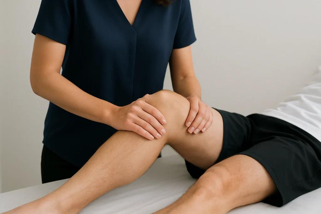
How are LCL Tears/Sprains/Strains Diagnosed?
A clinician will take a history of the injury and previous knee concerns and then perform a detailed physical examination, assessing:
- Localised tenderness along the outer knee.
- Swelling and bruising.
- Varus stress test to check for laxity.
- Evaluation of gait and range of motion.
- Assessment for associated injuries or nerve symptoms.
Which Tests Do Providers Use to Diagnose LCL Injuries?
Varus Stress Test
This is the primary clinical test for assessing LCL integrity. The examiner applies an inward (varus) force to the knee while it is bent to 0° and then to 30° of flexion.
- At 30°: Isolates the LCL by relaxing other stabilisers.
- At 0°: A positive test may suggest more extensive injury involving other structures (e.g. cruciate ligaments or capsule).
- Increased laxity or pain compared to the uninjured side may indicate an LCL tear.
External Rotation Recurvatum Test
Used to detect posterolateral rotatory instability.
The patient lies on their back, and the examiner lifts both legs by the big toes. Excessive hyperextension, varus alignment, or outward rotation of one leg compared to the other suggests injury to the posterolateral corner, which often includes the LCL.
Posterolateral Drawer Test
Performed with the knee flexed to 80–90°, the foot externally rotated, and the tibia pushed posteriorly.
This test helps detect instability or injury to the posterolateral corner and LCL by comparing side-to-side movement between knees.
Dial Test
Assesses external rotation of the tibia at 30° and 90° of knee flexion.
- Increased rotation at 30° only suggests isolated posterolateral corner injury.
- Increased rotation at both 30° and 90° suggests combined injury with the PCL.
MRI (Magnetic Resonance Imaging)
The gold standard for visualising soft tissue injuries. It provides detailed images of the LCL, surrounding ligaments, menisci, cartilage, and associated posterolateral structures.
It also helps assess whether the injury is partial or complete and detect additional damage.
X-rays
Used to rule out fractures, joint dislocations, or avulsion injuries where a piece of bone is pulled off by the ligament.
Weight-bearing or stress views can sometimes show subtle signs of instability.
Ultrasound
Though less commonly used, ultrasound may help visualise superficial ligament injuries, especially in children or thin patients.
It is a dynamic and accessible tool, but operator-dependent and less comprehensive than MRI.
Stress Radiographs
These are X-rays taken while the knee is under varus stress. They can quantify the degree of laxity and are sometimes used in surgical planning or for objective documentation of instability.
Specific Considerations for LCL Injuries
Isolated vs. Complex Injuries
Most LCL injuries occur in conjunction with other posterolateral structures. Isolated LCL tears are uncommon and tend to occur in lower-grade trauma. Complex injuries often involve the cruciate ligaments, menisci, and posterolateral corner, requiring surgical attention and longer rehabilitation.
Acute vs. Chronic Injuries
- Acute injuries: Easier to repair surgically when tissue quality is good.
- Chronic injuries: May lead to adaptive changes in gait and muscle use; reconstruction is often necessary, sometimes with staged procedures.
Associated Neurovascular Considerations
The common peroneal nerve lies just behind the LCL. Injury may present with foot drop, numbness along the shin or foot, or muscle weakness. Early diagnosis is crucial to prevent permanent deficits.
It is important to seek medical care from practitioners skilled in treating orthopaedic injuries. This knowledge ensures all factors are considered when forming a diagnosis and treatment plan.
How are LCL Tears/Sprains/Strains Treated?
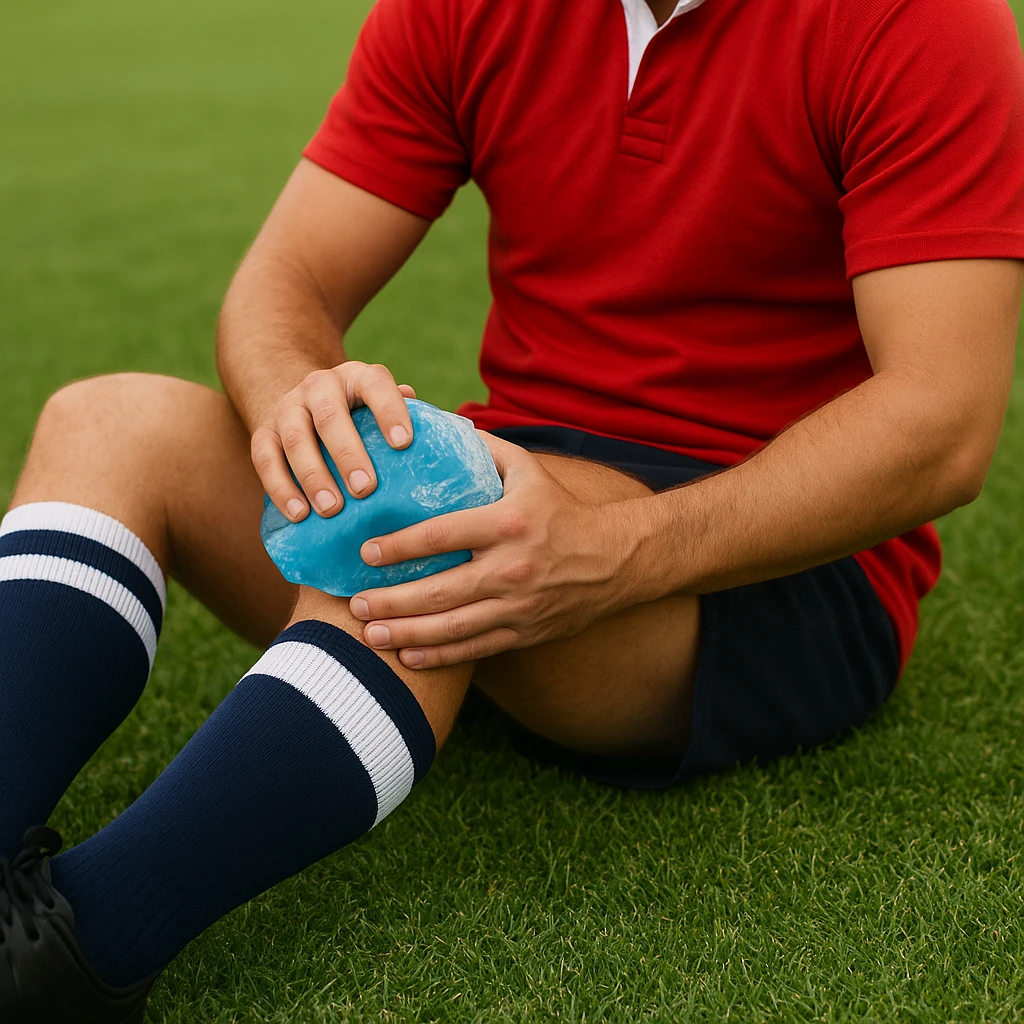
Non-Surgical Treatment (Grades 1–2):
- RICE protocol: Reduces swelling and inflammation in the acute phase.
- Hinged knee brace: Maintains alignment while allowing controlled motion.
- Physiotherapy: Focuses on restoring strength, balance, and flexibility.
- Lateral stability exercises: Side steps, resistance bands, proprioception drills.
- Anti-inflammatory medication: For pain control and swelling reduction.
- Gradual return to activity: Guided by symptom resolution and functional goals.
Surgical Treatment (Grade 3 or Complex Injuries):
- Primary repair: Best for acute, isolated tears with good-quality tissue.
- LCL reconstruction: Autograft or allograft is used when primary repair is not possible.
- Posterolateral corner reconstruction: Necessary for multi-ligament injuries.
- Addressing associated injuries: ACL, PCL, and meniscus tears are treated simultaneously.
- Postoperative rehabilitation: Tailored program for gradual recovery over months.
How Can I Prevent LCL Injuries?
The following techniques and ideas can help prevent LCL and other knee ligament injuries.
- Proper conditioning: Emphasise hamstring, quadriceps, and gluteal strength.
- Lateral and balance training: Improve neuromuscular control and reduce fall risk.
- Core stability: Reduces load on knees during dynamic activity.
- Neuromuscular training: Enhances joint position awareness and movement patterns.
- Correct technique: Important during cutting, jumping, and tackling.
- Protective equipment: Especially in contact sports.
- Recovery and rest: Avoid overtraining and ensure muscle recovery.
What Can I Expect if I Have an LCL Injury?
Most individuals with isolated Grade 1–2 injuries recover fully with non-operative care. Grade 3 injuries may require surgery and extensive rehabilitation, especially when associated with other ligament damage. Prompt diagnosis and appropriate treatment generally lead to good outcomes. Access Ortho offers rapid appointments for knee ligament injuries. The medical staff will guide you in your expected recovery timeline.
What is the Recovery Time from an LCL Injury?
- Grade 1: 2–4 weeks with rest and physiotherapy.
- Grade 2: 4–8 weeks, often with bracing and rehab.
- Grade 3: 6–9 months for full surgical recovery.
- Return to sport: Based on regaining strength, joint stability, and athlete confidence.
When Should I See a Specialist?
For knee ligament injuries, it is important to get advice from an orthopaedic specialist, as this is their area of expertise. Access Ortho offers rapid access to orthopaedic surgeons, ensuring you get the correct advice promptly. This will minimise time to diagnosis, ensure a correct diagnosis, and allow you to start treatment as soon as possible.
When Should I Visit a Fracture Clinic, such as Access Ortho?
Seek expert care if you experience:
- Outer knee pain: Especially after trauma.
- Swelling and bruising: On the outer knee.
- Instability: When changing direction or on uneven ground.
- Weight-bearing difficulty: Limping or inability to walk normally.
- Popping sensation: At the time of injury.
- Nerve symptoms: Numbness or tingling below the knee.
What is a Fracture Clinic?
Despite the name, fracture clinics, like Access Ortho, specialise in diagnosing and treating musculoskeletal injuries, including ligament sprains and tears. At Access Ortho, patients receive:
- Rapid specialist appointments
- Referral and review of advanced imaging (X-ray, MRI)
- Accurate diagnosis and timely treatment
- Referral to physiotherapy and rehab planning
- Personalised care without long ED wait times
Frequently Asked Questions
Why are LCL tears less common than MCL tears?
The LCL is located on the knee’s outer (lateral) side and is less exposed to direct forces. In most daily movements and sports, the knee is more likely to be pushed inward (placing stress on the MCL) than outward (which stresses the LCL), making MCL injuries more frequent.
Can an LCL tear heal without surgery?
Yes, most Grade 1 and Grade 2 LCL injuries heal well with non-surgical treatment such as rest, bracing, and physiotherapy. However, Grade 3 injuries or injuries involving other structures often require surgical repair or reconstruction.
How do I know if I’ve injured my LCL?
Typical signs include pain on the outer side of the knee, swelling, tenderness, and a feeling of instability, especially when pivoting or performing side-to-side movements. A clinical assessment and imaging are required for a definitive diagnosis.
What’s the difference between LCL and MCL injuries?
- The LCL stabilises the outer side of the knee and resists forces that push the knee inward.
- The MCL stabilises the inner side and resists forces that push the knee outward.
- The cause of injury, symptoms, and rehabilitation may differ depending on which ligament is affected.
Will I need crutches for an LCL sprain?
Crutches may be recommended initially if there is significant pain or instability, especially in Grade 2 or 3 injuries. They help offload weight and protect the healing ligament during the early stages.
How long should I wear a brace after an LCL injury?
Brace use typically lasts 2–6 weeks, depending on the severity. Your orthopaedic medical team will decide if you need a brace.
- Grade 1 injuries may only require bracing for a short period.
- Grade 2–3 injuries often need a hinged knee brace to maintain stability during recovery and physiotherapy.
Can I still play sports with a mild LCL sprain?
With a Grade 1 sprain, return to sport may be possible once pain subsides and stability is regained. It’s essential to follow a structured rehab plan and obtain medical clearance before returning to full activity.
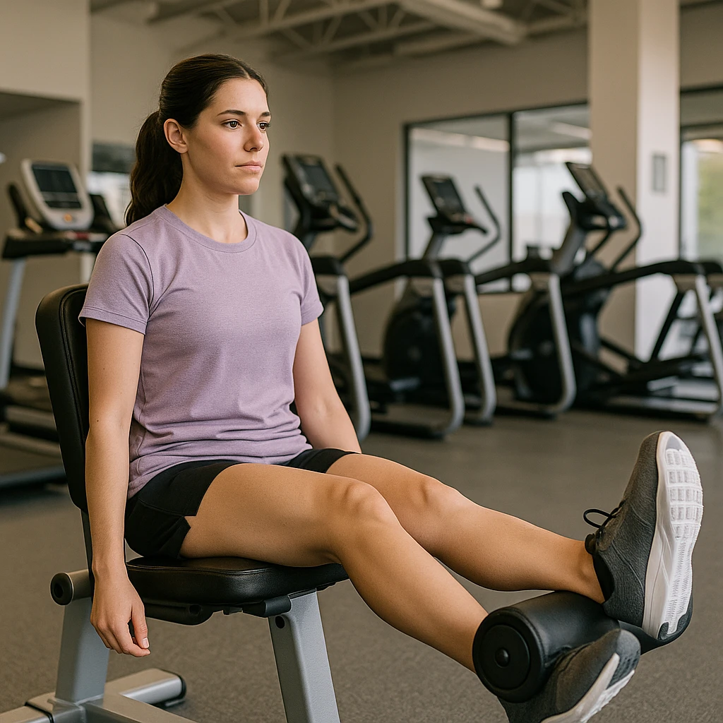
What exercises are best for LCL rehabilitation?
Key exercises include:
- Quad and hamstring strengthening
- Lateral step-ups and side lunges
- Balance and proprioception training (e.g. single-leg stands)
- Resistance band exercises targeting lateral stability
- Always start under the guidance of a physiotherapist.
Is surgery always necessary for a complete LCL tear?
Not always. Isolated Grade 3 LCL tears in low-demand individuals may be treated conservatively. However, surgery is generally recommended for complete tears, particularly in active patients or when other ligaments are involved.
Will an LCL injury cause long-term knee problems?
Most LCL injuries heal well with proper treatment. However, if untreated or poorly rehabilitated, they can lead to chronic instability, difficulty with lateral movements, and early joint degeneration.
How is a posterolateral corner injury different from a simple LCL tear?
A posterolateral corner injury involves multiple structures, such as the LCL, popliteus tendon, and popliteofibular ligament. These injuries cause rotational instability and are more complex than isolated LCL tears, often requiring surgery and longer rehabilitation.
What activities should I avoid during LCL recovery?
Avoid:
- Pivoting or cutting movements
- Lateral lunges and squats (early phase)
- Jumping or high-impact activities
- Sports drills until cleared by a clinician
Gradual reintroduction of activity is important in preventing re-injury.
Can physiotherapy alone heal an LCL tear?
Yes, physiotherapy is highly effective for Grade 1 and 2 injuries. It helps restore strength, mobility, and joint control. For partial or isolated tears, structured rehab can lead to full recovery without surgery.
How successful is LCL reconstruction surgery?
Surgical outcomes are generally positive when performed by an experienced orthopaedic surgeon, especially in acute or well-selected chronic cases. Success depends on injury complexity, surgical technique, and adherence to rehab.
Will I develop arthritis after an LCL injury?
An isolated LCL injury does not always mean you will get arthritis, but if instability persists or other structures are involved, there is an increased risk of joint wear and early-onset osteoarthritis. Timely treatment helps reduce this risk.