What is a Tibial Plateau Fracture?
A tibial plateau fracture is when there is a break in the bone in the upper part of the tibia (shin bone) that forms the knee joint. This type of fracture affects the flat upper surface of the tibia, known as the tibial plateau, which connects to the femur (thigh bone) and the fibula. The tibial plateau plays a crucial role in the stability of the knee joint, so fractures in this area can disrupt normal knee function.

Types of Tibial Plateau Fractures
Tibial plateau fractures are classified into different types based on the Schatzker classification system, which considers the pattern and severity of the fracture. This system helps guide treatment decisions and predict outcomes. There are six main types:
1. Schatzker Type I: Lateral Split Fracture
- Description: A simple fracture of the lateral tibial plateau (outer part of the knee) without significant depression of the bone surface.
- Common Cause: Typically seen in younger patients with strong bone, caused by valgus (inward) stress and axial loading.
- Treatment: Often managed with conservative treatment unless the fracture is displaced.
2. Schatzker Type II: Lateral Split-Depression Fracture
- Description: A combination of a split fracture and a depression of the lateral tibial plateau.
- Common Cause: High-energy trauma, often involving a forceful blow to the outer side of the knee.
- Treatment: Frequently requires surgical intervention to restore the joint surface.
3. Schatzker Type III: Lateral Depression Fracture
- Description: A pure depression fracture of the lateral tibial plateau without a split.
- Common Cause: This is seen in older patients with weaker bones, often from a low-energy fall.
- Treatment: This may require bone grafting and fixation, especially if the depression affects joint stability.
4. Schatzker Type IV: Medial Plateau Fracture
- Description: A fracture of the medial tibial plateau (inner part of the knee), often involving displacement or depression.
- Common Cause: High-energy trauma causing varus (outward) stress, often associated with ligament injuries.
- Treatment: Surgery is typically needed due to the potential for instability and misalignment.
5. Schatzker Type V: Bicondylar Fracture
- Description: Fractures involving both the medial and lateral tibial plateaus.
- Common Cause: High-impact trauma such as motor vehicle accidents or falls from a height.
- Treatment: Requires surgical fixation to stabilise the knee and restore alignment.
6. Schatzker Type VI: Complex Fracture with Metaphyseal-Disphyseal Dissociation
- Description: A severe fracture where the tibial plateau is broken into multiple fragments and separated from the shaft of the tibia.
- Common Cause: High-energy trauma such as car accidents or severe falls.
- Treatment: Requires extensive surgical reconstruction and may involve external fixation initially, followed by internal fixation.
Summary
The type of fracture determines the treatment approach and prognosis. For all tibial plateau fractures, early diagnosis and appropriate management are critical to prevent complications like post-traumatic arthritis, joint instability, or chronic pain. Access Ortho offers rapid appointments to ensure patients get specialist orthopaedic advice for their injury/injuries promptly.
How Common Are Tibial Plateau Fractures?
Tibial plateau fractures are relatively uncommon; they account for about 1% of all fractures and 8% of fractures involving older people. These injuries are more frequent in certain populations and scenarios, such as:
Demographics and Risk Factors
- Age Groups:
- Younger individuals: Typically caused by high-energy trauma (e.g., car accidents, sports injuries).
- Older individuals: More common due to low-energy trauma (e.g., falls), especially in those with weakened or osteoporotic bones.
- Active Populations:
- Athletes involved in contact sports or activities requiring sudden changes in direction (e.g., football, skiing).
- Gender Differences:
- They tend to occur more frequently in men under 50 due to higher exposure to high-impact trauma.
- It is more common in women over 50, likely due to osteoporosis-related fragility.
Symptoms and Causes
The symptoms of a tibial plateau fracture vary depending on the severity and location of fracture, but common signs include:
1. Pain
- Severe pain in the knee or upper shin, especially immediately after injury.
- Pain often worsens with movement or when attempting to bear weight.
2. Swelling
- Significant swelling around the knee joint is common, sometimes accompanied by bruising.
- Swelling may extend to the lower leg.
3. Deformity
- Visible misalignment or deformity of the knee may occur in severe or displaced fractures.
4. Difficulty Bearing Weight
- Inability or extreme difficulty in putting weight on the affected leg.
- A feeling of instability in the knee when trying to stand.
5. Limited Range of Motion
- Stiffness and difficulty bending or fully straightening the knee.
- The knee may feel locked in one position.
6. Tenderness
- Tenderness to touch over the upper shin or around the knee.
7. Bruising and Skin Discoloration
- Bruising around the knee or shin area due to bleeding beneath the skin.
8. Numbness or Tingling
- In cases of severe trauma, nerve or vascular damage may occur, causing numbness, tingling, or coldness in the lower leg or foot.
What Causes Tibial Plateau Fractures?
Tibial plateau fractures occur due to forces that exceed the strength of the tibial bone, often from significant trauma. The primary causes can be grouped into high-energy and low-energy mechanisms:
High-Energy Causes
- Motor Vehicle Accidents
- It is one of the most common causes, especially in younger individuals.
- The force from a car crash often drives the femur into the tibial plateau, causing fractures.
- Sports Injuries
- High-impact sports like football, rugby, or skiing can result in fractures due to sudden twisting, collisions, or falls.
- Landing awkwardly during jumping activities is also a common cause.
- Falls from Height
- Falling from a significant height, such as from a ladder or building, can cause axial loading (force transmitted along the leg), leading to fractures in the tibial plateau.
- Direct Impact to the Knee
- A direct blow, such as from a hard object or collision, can fracture the tibial plateau.
Low-Energy Causes
- Twisting Injuries
- Sudden twisting of the knee, particularly with the foot fixed, can place stress on the tibial plateau and lead to fractures.
- Falls (in Elderly or Osteoporotic Individuals)
- In people with weakened bones, such as the elderly or those with osteoporosis, even low-energy falls can cause a fracture.
Risk Factors for Tibial Plateau Fractures
- High-Impact Sports Participation
- Activities like skiing, football, and rugby increase the risk of knee trauma.
- Osteoporosis
- Weak bones make individuals more susceptible to fractures, even from low-impact injuries.
- Advanced Age
- Age-related bone loss and a higher likelihood of falls contribute to fracture risk.
- Previous Knee Injuries
- Prior injuries can weaken the knee joint, making it more vulnerable to future fractures.
- Occupational Hazards
- Jobs involving heavy lifting, climbing, or frequent exposure to falls (e.g., construction) increase risk.
- Motor Vehicle Use
- Accidents are a major cause of high-energy trauma leading to tibial plateau fractures.
Addressing these risk factors, such as strengthening bones or using protective gear, can help reduce the likelihood of fractures.
Complications of Tibial Plateau Fractures
- Post-Traumatic Arthritis
- A Tibial Plateau Fracture damages the joint surface, which can lead to long-term arthritis, which may cause pain, stiffness, and reduced mobility.
- Knee Instability
- Injury to ligaments or poor healing of the fracture can result in joint instability, affecting walking and daily activities.
- Malalignment
- Improper healing or inadequate reduction of the fracture may lead to deformities in the leg’s alignment, affecting function and biomechanics.
- Infection
- Open fractures or surgical interventions carry a risk of infection, which can complicate recovery.
- Compartment Syndrome
- Swelling and increased pressure within the leg’s compartments can compromise blood flow and nerve function, requiring emergency treatment.
- Nonunion or Delayed Union
- The fracture may fail to heal (nonunion) or take longer than expected to heal (delayed union), leading to prolonged disability.
- Vascular Complications
- Severe trauma may damage blood vessels around the knee, causing impaired circulation and tissue damage.
- Nerve Injury
- High-energy fractures can result in damage to nerves, causing numbness, tingling, or weakness in the lower leg or foot.
Early diagnosis, appropriate treatment, and regular monitoring are essential to minimise these complications and ensure optimal recovery. Access Ortho is a dedicated private fracture clinic specialising in fractures, sprains and strains. Access Ortho offers rapid appointments and specialist orthopaedic advice.
DIAGNOSIS AND TESTS
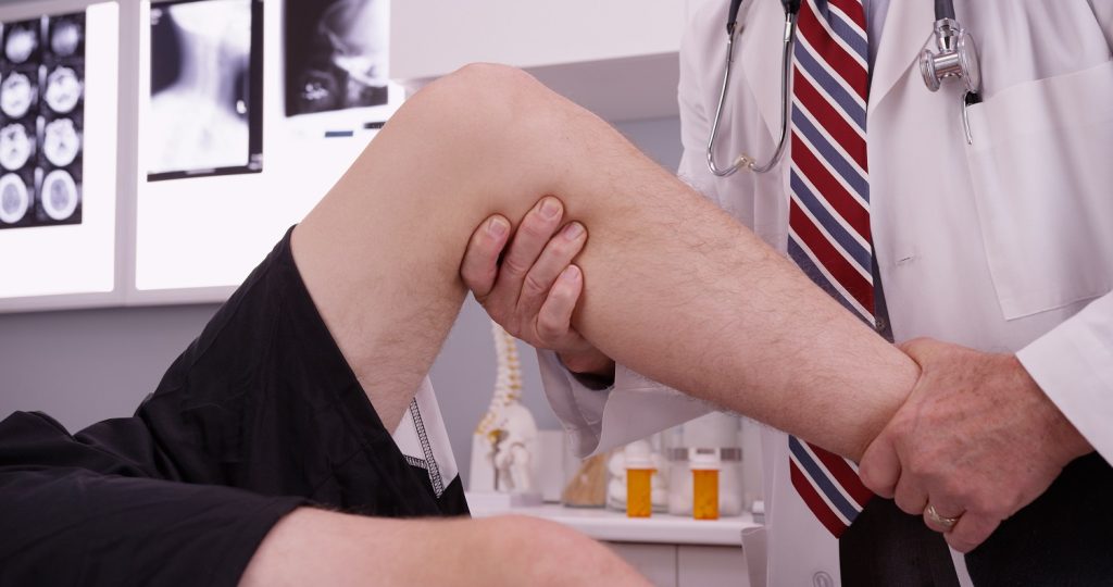
How Are Tibial Plateau Fractures Diagnosed?
Diagnosis begins with a thorough initial assessment, including:
- Medical History:
- Details about the injury mechanism (e.g., trauma, fall, or sports incident).
- Symptoms such as pain, swelling, inability to bear weight or numbness.
- Physical Examination:
- Inspection: Swelling, bruising, or deformity around the knee.
- Palpation: Tenderness over the tibial plateau.
- Range of Motion: Assessing knee stiffness or locking.
- Neurovascular Examination: Checking for blood flow (pulses) and nerve function in the lower leg and foot.
Diagnostic Tests for Tibial Plateau Fractures
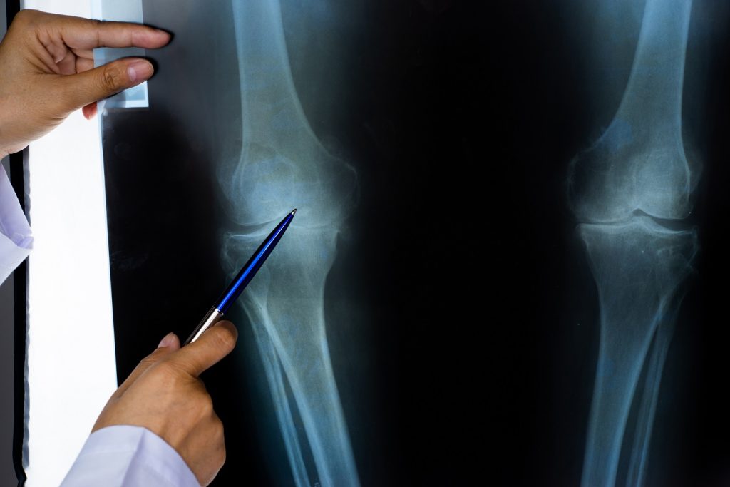
- X-rays:
- Initial imaging to detect fractures, assess alignment, and identify displacement.
- Standard views: Anteroposterior (AP), lateral, and oblique.
- CT Scans:
- Provides detailed images of the bone and fracture patterns.
- Useful for surgical planning in complex fractures.
- MRI Scans:
- Evaluates associated soft tissue injuries, such as ligament tears or meniscal damage.
- It also detects bone bruising or subtle fractures not visible on X-rays.
- Stress Radiographs:
- Performed under controlled conditions to assess ligament stability and knee alignment.
- Arteriography:
- Used when there’s suspicion of vascular injury, especially with high-energy trauma or displaced fractures.
- Compartment Pressure Measurements:
- Performed if compartment syndrome is suspected. Elevated pressure may indicate a need for urgent fasciotomy.
Special Considerations
- Associated Ligament Injuries:
- Common with tibial plateau fractures, they are often seen in conjunction with an anterior cruciate ligament or medial collateral ligament tear (ACL and MCL).
- It may require repair or reconstruction during fracture treatment.
- Meniscal Damage:
- Menisci can be torn or compressed during the injury. Addressing this is critical to restoring knee function and preventing arthritis.
- Articular Surface Involvement:
- Fractures involving the joint surface require precise reduction to prevent post-traumatic arthritis.
- Elderly Patients:
- Often, they have weaker bones, making fixation challenging. Conservative treatments are preferred in low-demand individuals.
- Athletes:
- Require optimal joint restoration to regain performance levels. Special attention is given to ligament and cartilage integrity.
- Complex Fracture Patterns:
- Bicondylar or comminuted fractures may require extensive surgical reconstruction and longer recovery times.
- Vascular Status:
- Monitoring for vascular injuries is essential to prevent complications such as tissue necrosis or amputation.
Tibial plateau fractures demand a multifaceted diagnostic approach, considering both bone and soft tissue injuries. Proper imaging and early detection of associated injuries or complications are crucial for optimising outcomes, particularly in high-demand individuals or those with complex fracture patterns. It is important to get specialist orthopaedic advice when assessing potential tibial plateau fractures. Access Ortho is led by orthopaedic surgeons, ensuring all patients get a high level of orthopaedic care and advice.
Management and Treatment
How Are Tibial Plateau Fractures Treated?
Treatment for tibial plateau fractures depends on the fracture type, severity, patient factors (age, activity level), and associated injuries. The primary goals are to restore the anatomy, ensure joint stability, and optimise knee function.
1. Emergency Care
- Stabilisation: Immobilise the leg using a splint or brace to prevent further injury.
- Pain Management: Administer pain relief medications or nerve blocks.
- Neurovascular Assessment: Evaluate circulation and nerve function to identify vascular compromise or compartment syndrome.
- Urgent Surgical Intervention: If vascular damage or compartment syndrome is present, immediate surgery is required.
2. Conservative management
For non-displaced or minimally displaced fractures:
- Immobilisation: Use of a knee brace or cast to limit movement and allow natural healing.
- Non-weight Bearing: Patients are advised to avoid weight-bearing on the affected leg for 6–8 weeks.
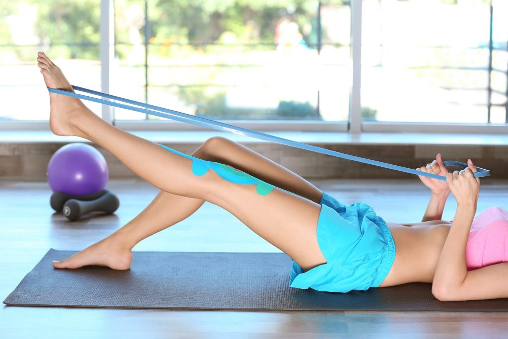
- Physiotherapy: Gradual rehabilitation begins after initial healing to restore range of motion and strength.
3. Surgical Interventions
Surgery is typically required for displaced, unstable, or complex fractures involving the joint surface.
Internal Fixation
- Metal plates and screws are used to realign and stabilise the fractured bone fragments.
- Commonly used for Schatzker Type II-VI fractures.
External Fixation
- An external frame stabilises the bone while soft tissues heal.
- Often used temporarily for severely comminuted fractures or in cases with soft tissue swelling.
Minimally Invasive Techniques
- Percutaneous Screw Fixation: Small incisions are used to insert screws for stabilisation.
- Advantage: Minimises soft tissue damage and promotes faster recovery.
Arthroscopy-Assisted Reduction
- A minimally invasive where a small camera and instruments are used to assess and treat fractures.
- Helps repair associated ligament or meniscal injuries.
- It is beneficial for fractures with joint depression or articular surface involvement.
4. Pain Management
- Medications: NSAIDs, paracetamol, or opioids for short-term use.
- Local Anaesthetics: Nerve blocks or intra-articular injections to manage acute pain.
5. Rehabilitation Protocols (general guide only)
- Early Phase (0–6 weeks): Focus on pain control, swelling reduction, and non-weight-bearing range of motion exercises.
- Intermediate Phase (6–12 weeks): Gradual weight-bearing and strength-building exercises under physiotherapy supervision.
- Late Phase (3–6 months): Full weight-bearing, advanced strengthening, and proprioception training to restore knee function.
- Return to Activity: High-demand activities or sports typically resume after 6–12 months.
Individualised care:
- Treatment plans must address associated injuries (e.g., ligament tears, meniscal damage) to ensure optimal outcomes.
- Regular follow-ups and imaging are necessary to monitor healing and prevent complications such as malalignment or arthritis.
By tailoring the treatment to the fracture and patient needs, Access Ortho helps ensure patients have the best functional recovery and long-term knee health possible.
Prevention
How Can I Prevent Tibial Plateau Fractures?
- Proper Protective Equipment: Use appropriate gear during high-risk sports to protect the knees.
- Sport-Specific Training: Strengthen muscles, improve balance, and practice safe techniques to reduce injury risks.
- Fall Prevention: Install handrails, use non-slip mats, and maintain suitable footwear to prevent falls, especially in older adults.
- Vehicle Safety: Wear seat belts and follow traffic regulations to minimise trauma during accidents.
- Workplace Safety: Follow safety protocols and use protective gear in hazardous jobs.
- Bone Health Maintenance: Maintain strong bones through an adequate diet, regular exercise, and addressing conditions like osteoporosis.
Outlook / Prognosis
Outlook / Prognosis: What to Expect with Tibial Plateau Fractures
The prognosis for tibial plateau fractures depends on the severity of the fracture, the effectiveness of treatment, and the patient’s adherence to rehabilitation. While many patients recover well, complex fractures may result in long-term challenges, such as joint stiffness or arthritis.
Recovery Timeline (This is a general guide only. It is important to be guided by your medical team)
1. Immediate Post-Injury Phase (0–2 Weeks)
- Focus: Stabilisation, pain management, and initial healing.
- Treatment may involve immobilisation with a brace or cast for non-surgical cases or surgical intervention for complex fractures.
- Swelling and pain are common, requiring rest, elevation, and medications.
2. Post-Operative Recovery (2–6 Weeks)
- Focus: Healing of bone and soft tissues.
- Patients typically remain non-weight-bearing during this phase.
- Stitches or staples are removed, and imaging is done to assess healing progress.
3. Rehabilitation Stages (6 Weeks–6 Months)
- Phase 1 (6–12 Weeks): Gradual range-of-motion exercises and partial weight-bearing as healing permits.
- Phase 2 (3–6 Months): Strengthening exercises, balance training, and functional activities to restore mobility and stability.
4. Return to Weight-Bearing (3–4 Months)
- Weight-bearing progresses based on fracture stability and radiographic evidence of healing.
- Full weight-bearing may be delayed for complex fractures or in older patients with poor bone quality.
5. Long-Term Outcomes (6 Months–1 Year or More)
- Most patients regain functional mobility within 6–12 months.
- Full recovery may take longer for severe fractures or high-demand activities.
- Long-term complications, such as post-traumatic arthritis or knee instability, may occur in some cases.
Expected Outcomes
- Favourable Outcomes: Non-displaced fractures or properly treated cases typically result in good functional recovery with minimal long-term issues.
- Challenging Cases: Complex or untreated fractures may lead to chronic pain, reduced mobility, or arthritis.
- Athletes: With proper rehabilitation, many athletes return to pre-injury levels of performance, though this may take up to a year.
Adhering to a structured treatment plan is essential for optimal recovery. Regular follow-ups and addressing complications promptly can improve long-term outcomes and minimise risks of chronic issues.
Fracture Clinic Information
When Should I Go to a Fracture Clinic?
Seek care from a fracture clinic such as Access Ortho if you experience fractures, sprains, or dislocations that require immediate attention, especially when you suspect a bone is broken, there is severe pain, or when the injury impairs mobility. Following an injury, you can attend Access Ortho without a referral.
Access Ortho offers quicker access to orthopaedics specialists compared to emergency rooms, providing faster diagnosis and treatment.
If you have already visited an emergency department or your GP, you can attend Access Ortho for follow-up care or a second opinion. Access Ortho is an affordable clinic that ensures rapid access to specialist Orthopaedic Care.
What is a Fracture Clinic?
A fracture clinic is a specialised medical facility dedicated to treating musculoskeletal injuries, including fractures, dislocations, and complex soft tissue injuries. Services available at Access Ortho include full assessment and diagnosis of upper and lower limb injuries, referral and follow-up of imaging such as X-rays and other radiology scans, non-invasive treatments like casting, surgical options, rehabilitation advice, and follow-up care.
At Access Ortho, you are getting care by medical staff specialised in Orthopaedics.
Frequently Asked Questions
How long does a tibial plateau fracture take to heal?
Healing time for a tibial plateau fracture typically ranges from 3 to 4 months. This will depend on the severity of the fracture and the treatment approach. Younger individuals may experience faster recovery, while older patients or those with conditions like osteoporosis may take longer.
When can I start putting weight through my leg after a tibial plateau fracture?
The timing for weight-bearing depends on the type of fracture and the treatment method. Generally, if surgery is not needed, weight-bearing may begin after 4–8 weeks. For surgical cases, weight-bearing is often introduced gradually, starting with partial weight-bearing after 6–12 weeks, depending on healing progress.
Will I need surgery for my fracture?
Not all tibial plateau fractures require surgery. Non-displaced fractures may heal well with immobilisation and non-weight-bearing, while displaced or complex fractures typically require surgical intervention to restore alignment and joint stability.
How will surgery affect my knee function?
Surgery aims to restore the correct alignment of the knee, but you may experience some initial pain and swelling following the procedure. With proper rehabilitation, your knee function will improve over time, although full recovery may take several months. Some people may have lingering stiffness or weakness, especially if the joint surface was damaged.
What type of physiotherapy will I need?
Physiotherapy will focus on restoring range of motion, strengthening muscles, and improving balance and proprioception. The rehabilitation process will begin after initial healing, with exercises progressing as tolerated, aimed at regaining function and mobility.
When can I return to sports or work?
Returning to sports or work depends on the severity of the fracture and the recovery process, along with the type of work and sport you wish to return to. For lighter activities, return may be possible after 1–4 months, while for high-impact sports, full recovery may take 6–12 months. Your healthcare provider will assess your individual progress and give specific guidelines based on your recovery.
Will I develop arthritis in my knee?
After a Tibial Plateau Fracture, there is a risk of developing post-traumatic arthritis, especially if the joint surface was affected or if there was significant displacement. Follow-ups with your doctor can help manage symptoms, and early intervention may help slow the progression of arthritis.
How long will I need to wear a brace or cast?
The duration of wearing a brace or cast depends on the fracture and treatment. Typically, a cast or brace is worn for 6–8 weeks in non-surgical cases. If surgery is required, a brace may be needed for 6–12 weeks, depending on how well the bone heals.
Can I drive with a tibial plateau fracture?
You should not drive while recovering from a tibial plateau fracture. You should talk to your doctor to determine when it is safe for you to resume driving.
What activities should I avoid during recovery?
During recovery, you should avoid high-impact activities like running, jumping, or heavy lifting that could strain the knee. Additionally, activities that may risk re-injury, such as sports or walking uneven terrain, should be avoided until your doctor clears you to return to normal activities.
How can I manage pain and swelling?
To manage pain and swelling, elevate the injured leg, apply ice packs, and take pain medications as prescribed. Following your doctor’s instructions on rest and activity levels is crucial for managing these symptoms effectively.
What signs indicate possible complications?
If you experience increased pain, swelling, signs of infection (like redness, warmth, or fever), numbness, tingling in your foot, or an inability to move your toes, these could be signs of complications. It’s important to seek medical attention promptly if any of these occur to ensure proper management and avoid further issues.