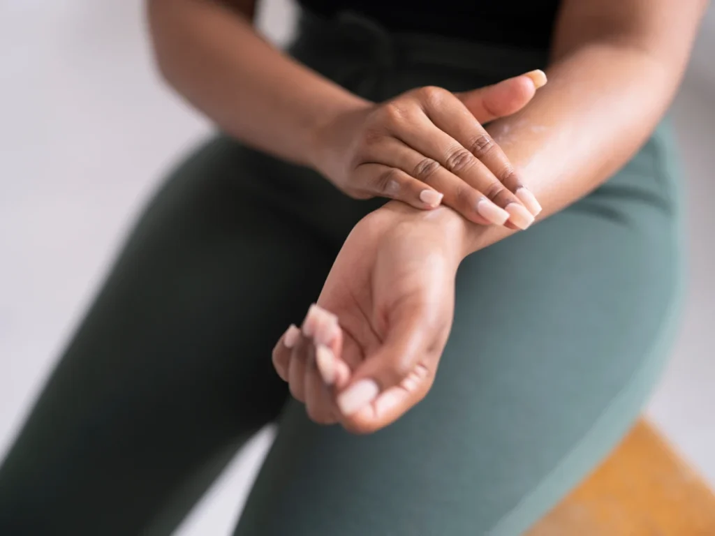
Definition of an Ulnar Fracture
An ulnar fracture refers to a break in the ulna, one of the two bones in the forearm, the other being the radius. It can occur as a result of direct trauma (e.g., a fall or impact) or repetitive stress. Depending on the location and severity, an ulnar fracture can affect the forearm’s functionality and may involve other nearby structures such as muscles, nerves, or the radius.
Anatomy of the Ulna and Its Role in the Forearm
The ulna is one of the two long bones in the forearm and is located on the side opposite the thumb (medial side). It runs parallel to the radius, forming the skeleton of the forearm. Key features of the ulna include:
- Proximal end (near the elbow): The olecranon process forms the bony point of the elbow and provides attachment for the triceps muscle.
- Distal end (near the wrist): The ulna tapers and ends in the head of the ulna, with a small projection called the styloid process.
- Shaft: The long part of the ulna that runs along the length of the forearm.
The ulna’s main functions include:
- Support and stability of the forearm: It works with the radius to allow for movements like pronation and supination (rotation of the forearm).
Articulation at the elbow and wrist: The ulna forms part of both the elbow and wrist joints, helping in elbow flexion and extension and contributing to wrist movement.

Types of Ulnar Fractures
There are different types of ulnar fractures based on the location and the mechanism of injury. Each type may require a different treatment approach.
Proximal Ulnar Fractures (Olecranon Fractures)
- Location: These fractures occur at the olecranon process, the bony prominence of the ulna that forms the point of the elbow.
- Mechanism of injury: This is commonly caused by a direct strike to the elbow or a fall onto a bent elbow.
- Symptoms: Pain, swelling, and inability to straighten the elbow.
- Treatment: Depending on the fracture, treatment may involve either immobilisation with a splint or cast or surgical repair using plates or screws.
Ulnar Shaft Fractures
- Location: These fractures occur along the mid-portion of the ulna’s shaft.
- Mechanism of injury: Often results from direct trauma to the forearm (e.g., blocking an object in a defensive position).
- Types: Ulnar shaft fractures can be simple (clean break) or complex (involving multiple bone fragments).
- Treatment: Most ulnar shaft fractures require immobilisation or surgery, depending on alignment and displacement.
Distal Ulnar Fractures
- Location: These occur near the wrist at the distal end of the ulna.
- Mechanism of injury: Often associated with fractures of the distal radius, such as Colles’ fracture, typically from a fall on an outstretched hand.
- Symptoms: Pain, swelling, and decreased wrist motion.
- Treatment: This may involve splinting, casting, or surgical fixation, especially if the radius is also fractured.
Monteggia Fractures (Ulnar Fracture with Radial Head Dislocation)
- Description: A Monteggia fracture is a fracture of the proximal ulna (usually the shaft) along with a dislocation of the radial head at the elbow.
- Mechanism of injury: This is typically caused by a fall onto a hand with the forearm rotated.
- Symptoms: Visible deformity, pain, swelling, and restricted elbow and forearm motion.
- Treatment: Surgical intervention may be required to repair the ulnar fracture and realign the radial head.
These different types of ulnar fractures each have specific treatments, but all involve some degree of rehabilitation to restore the function of the forearm and elbow.
How Common are Ulnar Fractures?
Ulnar fractures are relatively common, particularly when combined with other fractures (such as the radius) or in specific injury patterns like the Monteggia fracture. The incidence varies depending on age, gender, activity levels, and the nature of the trauma.
Incidence Rates
- General Incidence:
- Ulnar fractures account for approximately 5% of all fractures in adults.
- Combined with radius fractures: Ulnar fractures are often associated with radius fractures, making distal forearm fractures some of the most common fractures, especially in older adults. In developed countries, the incidence rate of distal forearm fractures is around 100-200 per 100,000 people annually.
- Fracture Specific Incidences:
- Isolated ulnar shaft fractures (also called “nightstick fractures”) are less common and occur more often as a result of direct trauma, like defensive injuries.
- Olecranon fractures (proximal ulna) account for about 10% of all upper extremity fractures, with an incidence rate of 10-20 per 100,000 people per year.
Demographics Most Affected by Ulnar Fractures
- Age:
- Younger individuals:
- Ulnar fractures, especially those caused by high-energy trauma (sports, motor vehicle accidents), are more common in younger adults, typically between 15 and 40 years old.
- Monteggia fractures are also more common in children, especially between 4 and 10 years.
- Older adults:
- Distal forearm fractures, often involving both the ulna and radius, are very common in people over the age of 50. These are typically low-energy injuries, such as falls from standing height, often linked to osteoporosis.
- Younger individuals:
- Gender:
- Women are more frequently affected by distal ulnar fractures, especially postmenopausal women, due to the higher incidence of osteoporosis, which weakens bones and makes fractures more likely.
- Men tend to experience isolated ulnar fractures and Monteggia fractures more frequently due to high-energy impacts from sports or manual labour.
- Activity Levels:
- Athletes and manual workers are at a higher risk of ulnar fractures due to their increased exposure to high-impact activities.
- Children are prone to ulnar fractures from falls during play, particularly at younger ages.
Symptoms and Causes
What Causes Ulnar Fractures?
Ulnar fractures can result from various mechanisms, depending on the type and location of the fracture. The most common causes include direct trauma, falls, and repetitive stress. Each mechanism of injury contributes to different types of ulnar fractures.
Direct Trauma to the Forearm
- Mechanism: A direct blow or impact to the forearm is a frequent cause of isolated ulnar fractures, often referred to as “nightstick fractures.” These fractures typically occur when the forearm is used to block or defend against an object, such as in cases of assault or contact sports.
- Injury Type: Direct trauma usually leads to ulnar shaft fractures, where the mid-portion of the ulna breaks. These can be simple (a clean break) or more complex (with multiple fragments).
- Common Scenarios:
- Defensive injuries (e.g., trying to block a blow).
- Accidents involving direct impact (e.g., car accidents, falls onto an object).
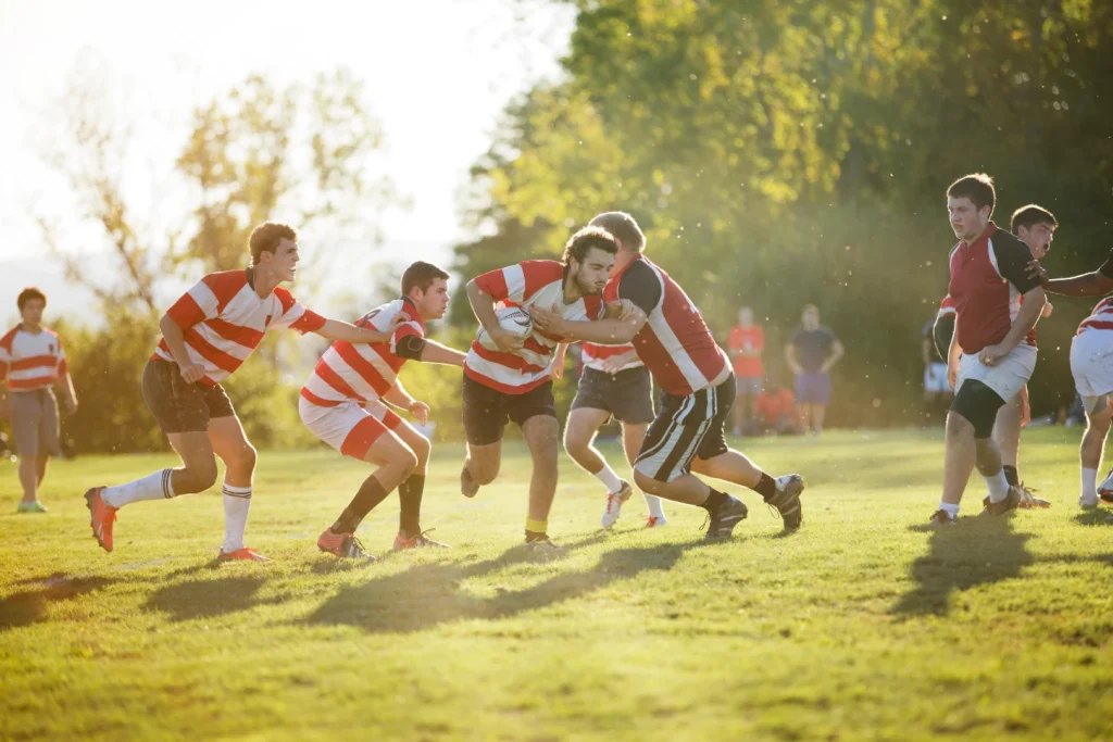
- Contact sports for example martial arts, football or rugby.
Fall onto an Outstretched Hand (FOOSH)
- Mechanism: Falling onto an outstretched hand is a common cause of fractures in the forearm. When an individual attempts to break their fall by extending the hand, the force of the impact is transmitted up the arm to the ulna and radius. This can result in fractures, especially when the bones are weakened (e.g., in osteoporosis).
- Injury Type:
- Distal ulnar fractures: These often occur alongside a distal radius fracture, particularly in older adults. A common example is a Colles’ fracture (fracture of the distal radius with dorsal displacement), which may also involve the ulna.
- Proximal ulnar (olecranon) fractures: These can occur if the elbow is flexed during the fall, causing the force to impact the proximal ulna.
- Common Scenarios:
- Slips and falls in everyday activities, especially in older adults.
- Falls during sports or physical activities.
Sports-Related Injuries
- Mechanism: High-energy impacts during sports activities can cause fractures in the ulna, particularly in sports or activities involving a high risk of falls. The forearm may suffer significant trauma either through direct contact or from the transmission of force during a fall.
- Injury Type:
- Monteggia fractures: These are common in sports where falls on the arm occur, such as gymnastics, skateboarding, or cycling. This injury involves a break in the proximal ulna along with a dislocation of the radial head.
- Olecranon fractures: Athletes may also experience fractures of the olecranon (the tip of the elbow) during falls or collisions.
- Common Scenarios:
- Gymnastics, cycling, and skateboarding (due to falls).
- Contact sports like rugby, football, and wrestling (due to impacts).
Stress Fractures in Athletes
- Mechanism: Stress fractures are caused by repetitive, prolonged loading on the bone, leading to microscopic fractures that can worsen over time if not treated. In athletes, particularly those who engage in weight-bearing or impact activities, repeated strain on the ulna can eventually lead to a stress fracture.
- Injury Type: Stress fractures typically affect the midshaft (shaft) of the ulna. These fractures may not be immediately symptomatic but gradually lead to increasing pain and tenderness in the forearm.
- Common Scenarios:
- Athletes involved in sports with repetitive arm motions (e.g., tennis, baseball, or gymnastics).
- Weightlifters or rowers, where repetitive strain is placed on the forearm bones.
What are ulnar fracture risk factors?
- Age-Related Risks
- Older adults: More prone to distal ulnar fractures, especially due to falls, as bones become more fragile with age.
- Children: Increased risk of ulnar fractures from falls and trauma during play or sports, such as Monteggia fractures.
- Osteoporosis and Bone Density Issues
- Osteoporosis weakens bones, this increases the risk of fractures from low-impact trauma. Postmenopausal women are particularly at risk of ulnar fractures associated with decreased bone density.
- Participation in High-Risk Sports or Activities
- Sports such as football, martial arts, gymnastics, and cycling increase the risk of ulnar fractures due to high-impact collisions, falls, or repetitive strain.
- Occupational Hazards
- Jobs involving manual labour, repetitive motions, or high physical risk (e.g., construction work or heavy lifting) increase the likelihood of direct trauma or stress fractures in the ulna.
These factors contribute to the likelihood of experiencing an ulnar fracture due to trauma, falls, or stress on the bone.
What are the complications of ulnar fractures?
- Nonunion or Malunion
- Nonunion: The fractured bone fails to heal, requiring prolonged treatment or surgery.
- Malunion: The bone heals improperly, leading to deformities or impaired function.
- Nerve Damage (Particularly the Ulnar Nerve)
- Fractures, especially near the elbow, may damage the ulnar nerve, resulting in numbness, tingling, or weakness in the hand and fingers.
- Compartment Syndrome
- Swelling and increased pressure in the forearm muscles can restrict blood flow, potentially leading to tissue damage, requiring emergency intervention to prevent permanent loss of function.
- Impaired Forearm Rotation and Grip Strength
- Improper healing or stiffness after an ulnar fracture can reduce the ability to rotate the forearm (pronation and supination) and weaken grip strength, affecting daily activities and overall arm function.
These complications can affect long-term recovery and may require additional treatments or surgery to restore function. It is important to seek orthopaedic advice for Ulnar fractures. Access Ortho specialises in fracture care.
Diagnosis and Tests
How are ulnar fractures diagnosed?
Diagnosing ulnar fractures involves a combination of clinical examination, assessing nerve and blood supply, and various imaging tests to determine the fracture’s location, severity, and associated injuries.
Clinical Examination Techniques
- Physical inspection: The medical practitioner checks for visible deformities, swelling, bruising, or open wounds that may suggest a fracture.
- Palpation: Gentle pressure is applied along the ulna to locate areas of tenderness or instability.
- Range of motion assessment: Testing the ability to move the wrist, elbow, and forearm helps determine if the fracture is affecting joint function.
- Functional testing: To check for impaired movement, the patient may be asked to make a fist, grip objects, or rotate the forearm.
Assessing Nerve Function and Blood Supply
- Nerve examination: The ulnar nerve may be evaluated for numbness, tingling, or weakness, especially in the ring and little fingers. Doctors may perform sensory tests to ensure nerve integrity.
- Circulatory assessment: Checking the pulse and capillary refill in the fingers helps assess the blood supply to the forearm and hand. Any compromise in circulation could indicate more serious complications like compartment syndrome.
Which tests do providers use to diagnose ulnar fractures?
X-rays for Ulnar Fractures
- Primary diagnostic tool: X-rays are the first-line imaging technique for ulnar fractures. They provide clear images of bone alignment and can identify most types of fractures (e.g., shaft, proximal, and distal fractures).
- Multiple views: X-rays are typically taken from multiple angles (anteroposterior, lateral) to assess the fracture from different perspectives.
CT Scans for Complex Fracture Patterns
- Detailed imaging: CT scans offer a more detailed view of complex fracture patterns, particularly in cases of multi-fragmented fractures or fractures involving joints (e.g., the olecranon or distal ulna).
- 3D reconstruction: CT images may be reconstructed into 3D models to assist in pre-surgical planning for intricate fractures.
MRI in Cases of Suspected Soft Tissue Damage
- Soft tissue evaluation: MRI is used if there is suspicion of damage to the ligaments, tendons, or nerves around the ulnar fracture.
- Associated injuries: MRI can also help assess injuries like muscle tears or joint capsule involvement that might accompany the fracture, particularly in cases where X-rays do not reveal significant damage.
Bone Scans for Stress Fractures
- Detecting stress fractures: Bone scans are useful when a stress fracture is suspected but may not be visible on X-rays, especially in athletes. The scan detects areas of increased bone activity (hot spots) that indicate a stress fracture.
- Early detection: Bone scans can identify fractures early in their development before they fully develop into visible breaks.
Access Ortho specialises in the assessment of fractures, including Ulnar fractures. This ensures an accurate diagnosis and a comprehensive treatment plan, minimising complications.
Specific Considerations
Ulnar Fractures in Children (Greenstick Fractures)
- Description: Children’s bones are softer and more flexible, making them prone to greenstick fractures, where the bone bends and cracks rather than breaking completely.
- Location: Often affects the midshaft of the ulna and may involve the radius as well.
- Treatment: Greenstick fractures typically heal well with immobilisation, such as casting. The bone’s natural ability to remodel in children usually leads to complete recovery without surgery.
- Prognosis: Children generally recover quickly from these fractures due to the rapid healing of young bones.
Ulnar Fractures in the Elderly
- Description: In older adults, fractures are often caused by low-energy trauma, such as a fall onto an outstretched hand (FOOSH). These fractures frequently occur in the distal forearm.
- Osteoporosis: Age-related bone density loss, particularly in postmenopausal women, increases the risk of ulnar fractures and complicates healing.
- Common Types:
- Distal ulnar fractures: Often occur with distal radius fractures (e.g., Colles’ fracture).
- Olecranon fractures: May result from a direct fall onto the elbow.
- Treatment: Casting may be required, but surgery (e.g., plating or pinning) is more common due to fragility and the likelihood of poor alignment during healing.
- Prognosis: Healing may be slower, and there is a higher risk of complications like nonunion or malunion.
Athletes and Ulnar Stress Fractures
- Description: Stress fractures occur due to repetitive strain or overuse, especially in athletes involved in sports requiring constant upper limb use (e.g., tennis, gymnastics, rowing).
- Location: Most commonly occurs in the midshaft of the ulna.
- Symptoms: Pain gradually increases over time, typically worsening with activity and improving with rest.
- Treatment: Rest and activity modification are essential to prevent the stress fracture from worsening. In more severe cases, immobilisation or surgery may be required.
- Prognosis: With proper rest and rehabilitation, stress fractures in athletes usually heal without long-term complications, though recurrence is possible if training resumes too soon.
Ulnar Fractures Associated with Radial Injuries
Ulnar fractures often occur in conjunction with radial fractures due to the close anatomical relationship between these two bones in the forearm. The most common injury pattern is a combined ulnar and radius fracture, such as in a Galeazzi fracture (distal radius fracture with distal ulnar dislocation).
- Monteggia Fractures: Involves a fracture of the proximal ulna along with a dislocation of the radial head. This injury requires prompt surgical intervention to realign the bones and restore normal joint function.
- Treatment: Most combined ulnar and radial fractures require surgical fixation (plates, screws) to ensure proper alignment and healing.
- Prognosis: Early treatment is key to preventing complications like malunion or limited range of motion in the forearm. The involvement of both bones can lead to a more complicated recovery and a longer rehabilitation process.
These specific considerations highlight the importance of age, activity level, and associated injuries when diagnosing and treating ulnar fractures. Each group has unique challenges, from managing fragile bones in older adults to treating stress fractures in athletes. Access Ortho are specialists in orthopaedic care.
Management and Treatment
How are Ulnar Fractures Treated?
Treatment for ulnar fractures depends on the severity, location, type of fracture, and the patient’s age and activity level. Management can range from conservative approaches to surgical interventions.
Conservative Management for Stable Fractures
- Indication: Conservative management is appropriate for stable fractures where the bone fragments are well-aligned and have minimal displacement.
- Examples:
- Greenstick fractures in children.
- Isolated ulnar shaft fractures with minimal displacement.
- Distal ulnar fractures without involvement of the radius or significant joint disruption.
- Treatment:
- Cast immobilisation: The forearm is immobilised in a cast to keep the bone stable and allow natural healing.
- Duration: Immobilisation typically lasts 4-6 weeks, depending on the fracture and healing progress.
- Follow-up: Regular X-rays are taken to ensure proper healing and alignment.

Surgical Interventions: Indications and Techniques
- Indications for Surgery:
- Displaced or unstable fractures: Fractures that have significant displacement or misalignment, such as those involving both the ulna and radius, often require surgery.
- Open fractures: This is when the bone breaks and pierces through the skin, surgery is necessary to clean the wound and stabilise the fracture.
- Comminuted fractures: In cases with multiple bone fragments, surgical fixation is usually required.
- Fractures associated with nerve or vascular damage, or injuries like Monteggia fractures, also require surgical repair.
- Surgical Techniques:
- Open reduction and internal fixation, which is known as ORIF: The surgeon repositions the bone fragments (open reduction) and uses metal plates, screws, or rods to stabilise the bone (internal fixation).
- Intramedullary nailing: This procedure involves a rod being inserted into the marrow canal of the ulna to hold the bone fragments together, often used in shaft fractures.
- External fixation: Sometimes used in severe fractures or when the skin and surrounding tissues are damaged. A frame outside the body holds the bones in position with pins inserted into the bone.
- Recovery: After surgery, a cast or splint may still be used to protect the repair. Rehabilitation to restore movement and strength is critical during the healing process.
Cast Immobilisation Techniques
- Short arm cast: This is typically used for distal ulnar fractures or stable fractures of the ulna. It immobilises the wrist and forearm but allows elbow movement. Access Ortho uses EXOS casts for immobilisation of some ulna fractures. These are thermoplastic moulded casts. Fibreglass, plaster, or a referral for a hand therapy splint may also be recommended.
- Long arm cast: Applied for proximal or shaft fractures. It immobilises the elbow, forearm, and wrist to restrict movement and ensure proper healing.
Pain Management Strategies
- Initial pain relief:
- Ice and elevation: Used immediately after injury to reduce swelling and pain.
- Immobilisation: Stabilising the fracture with a splint or cast can reduce pain by preventing movement at the fracture site.
- Medications:
- Nonsteroidal anti-inflammatory drugs (NSAIDs): Commonly used for mild to moderate pain relief and inflammation control.
- Panadol (paracetamol): Another option for managing pain without the anti-inflammatory effect.
As specialists in Orthopaedic Care, the Access Ortho team will assess your injury and explain the appropriate treatment required for a successful recovery. Casting can be done in the clinic. If you require surgery, they will help arrange this and provide a referral. Non-surgical follow-up care is in the Access Ortho clinic.
Prevention
How can I prevent ulnar fractures?
Proper Protective Gear in Sports
- Wearing protective gear, such as wrist guards, elbow pads, and forearm guards, can reduce the risk of ulnar fractures, especially in high-impact sports like football, skateboarding, or martial arts.
Fall Prevention Strategies
Minimising the risk of falls by ensuring safe environments (e.g., removing tripping hazards) and improving balance through exercises can prevent fractures, especially in older adults.
Bone Health and Nutrition
Maintaining strong bones through a diet rich in calcium and vitamin D helps reduce the risk of fractures. Regular check-ups to assess bone density, particularly for older adults, can identify and address bone health issues like osteoporosis.
Strengthening Exercises for Forearm Muscles
Engaging in forearm strengthening exercises improves muscle tone, supporting and protecting the bones during physical activities or in the event of a fall.
Outlook / Prognosis
What can I expect if I have an ulnar fracture?
Short-Term Recovery Milestones
- First few days: Swelling, pain, and limited movement in the affected arm. Immobilisation with a splint or cast is typically initiated immediately.
- 1-2 weeks: Initial reduction in pain and swelling. Follow-up X-rays may be taken to confirm that the bone alignment is proper.
- 4-6 weeks: Cast or splint may be removed for stable fractures. Pain continues to subside, and gentle range-of-motion exercises may begin under supervision.
- 6-12 weeks: Bone healing progresses. If surgery is required, this is the period where post-surgical rehabilitation intensifies.
Long-Term Prognosis and Functionality
- Restoration of function: Most patients regain full arm function, though recovery time varies depending on the severity and location of the fracture.
- Full strength recovery: It may take several months for the forearm’s strength to return to normal, especially in cases of more complex fractures or those requiring surgery.
- Potential complications: If complications arise (e.g., nonunion, nerve damage), they may impact long-term functionality. However, with appropriate treatment and rehabilitation, most people regain a high level of forearm strength and mobility.
What is the Recovery Time from an Ulnar Fracture?
Recovery time depends on the fracture type, treatment approach, and individual healing capacity.
Typical Healing Time Frames for Different Types of Ulnar Fractures
- Stable fractures (e.g., greenstick fractures in children): Healing generally occurs in 4-6 weeks with proper immobilisation.
- Displaced or complex fractures: Healing may take 8-12 weeks or longer, especially if surgery is required.
- Surgical repair: After surgery, bone healing typically takes 8-12 weeks, followed by several months of rehabilitation for full function recovery.
- Stress fractures: It may take 6-8 weeks of rest and rehabilitation for proper healing.
Factors Affecting Recovery Time
- Age: Younger individuals typically heal faster, while older adults may experience slower recovery due to bone density issues.
- Type of fracture: Simple fractures heal more quickly than complex, comminuted, or multi-fragmented fractures.
- Treatment type: Fractures treated conservatively (with a cast) tend to heal faster than those requiring surgery, which adds recovery time.
- Bone health: Conditions like osteoporosis or poor bone density can prolong healing.
- Post-treatment rehabilitation (if required): Adherence to physiotherapy and strength exercises will influence the speed of recovery and return to full functionality.
In summary, recovery from an ulnar fracture can take 4 weeks up to several months, depending on the fracture, the type of treatment required, and individual healing factors. With proper care and rehabilitation, most patients achieve full functional recovery.
When should I go to a fracture clinic?
Signs and Symptoms Requiring Immediate Medical Attention
You should seek medical attention at a fracture clinic such as Access Ortho if you experience any of the following:
- Severe pain: Intense, unmanageable pain that doesn’t improve with rest or over-the-counter pain medications.
- Visible deformity: A noticeable bend or misalignment in the forearm or elbow, indicating a possible fracture.
- Swelling and bruising: Significant swelling or bruising around the affected area, especially if it worsens over time.
- Inability to move the arm: Difficulty moving the wrist, elbow, or fingers, or inability to bear weight or use the arm.
- Open fracture: If the bone is protruding through the skin or there is an open wound, seek immediate care to prevent infection. If you have an open fracture, seek immediate care at the nearest hospital emergency department.
- Signs of nerve or vascular injury: Numbness, tingling, weakness in the hand or fingers, or changes in skin colour (pale, cool) in the fingers could indicate nerve or blood vessel damage.
Follow-Up Care and Monitoring
- Initial evaluation: After the initial diagnosis and treatment, follow-up visits to a fracture clinic are essential to monitor healing and ensure proper alignment of the fracture. Access Ortho will book follow-up appointments; it is important to attend these.
- Regular X-rays: Scheduled X-rays during follow-up visits will help assess bone healing and alignment, allowing for adjustments in treatment if necessary.
- Symptom management: Discuss any ongoing pain or functional limitations during follow-up visits to adjust pain management strategies or rehabilitation exercises.
- Complication monitoring: During follow-up appointments, it is crucial to observe for any signs of complications, such as nonunion, malunion, or nerve damage. If complications arise, immediate intervention may be necessary.
Access Ortho offers urgent care for musculoskeletal injuries such as fractures, sprains and strains.
What is a fracture clinic?
A fracture clinic such as Access Ortho offers specialist care in orthopaedic injuries such as fractures and sprains. A fracture clinic differs from Urgent Care or Emergency Departments as they only treat musculoskeletal injuries and are experts in managing these conditions. In addition, Access Ortho offers follow-up care in the clinic with orthopaedic surgeons. This ensures you receive specialist care until a full recovery is made.
At a Fracture Clinic such as Access Ortho, your injury will be assessed, medical history taken, and radiology arranged. Based on these findings and in collaboration with the orthopaedic surgeons, a diagnosis will be made. Non-surgical treatment and follow-up will occur at the clinic, and if necessary, a referral for surgery will be arranged.
Access Ortho offers Urgent Care for musculoskeletal injuries, and no referral is required to attend.
What to expect when you attend your fracture clinic appointment
A fracture clinic appointment is crucial for assessing the extent of your ulnar fracture and developing a tailored treatment plan. Here’s an overview of what typically happens during the visit.
Initial Assessment
Medical History:
- The medical practitioner will begin by taking a thorough medical history. They’ll ask about:
- How the injury occurred (e.g., fall, sports injury, etc.).
- Symptoms you’ve been experiencing, such as pain level, swelling, and wrist mobility.
- Previous medical conditions (e.g., osteoporosis) or any prior fractures or surgeries involving the wrist.
Physical Examination:
- The clinician will carefully assess the wrist, hand, and forearm, looking for:
- Swelling and bruising.
- Deformity or signs of bone displacement.
- Range of motion: You may be asked to try moving your fingers, wrist, and hand to check how much movement you have and to assess pain with movement.
- Circulation and Nerve Function: The doctor will check for normal blood flow (pulse in the wrist) and nerve function (sensation in the fingers and hand).
Diagnostic Imaging
If not already performed, diagnostic imaging is essential to diagnose and confirm the fracture type and severity. Access Ortho will refer for these. Radiology practices are located nearby so you can return to the clinic after your x-rays for the results.
Treatment Plan Discussion
The medical practitioner will discuss treatment options. Depending on the fracture type and severity, this can include conservative or surgical treatment. If surgery is required, Access Ortho will help arrange this.
Follow-Up Appointments
Before leaving the fracture clinic at Access Ortho, you’ll be scheduled for follow-up visits to monitor your healing. During these visits the Orthopaedic Surgeon will assess your progress, arrange check radiology if required, consider any complications and alter treatment plans if required. You will be able to ask any questions you may have. If at anytime during your follow-up care a referral is required for hand therapy this can be arranged.
Commonly Asked Questions
Can you move your arm with an ulnar fracture?
In many cases, movement may be limited due to pain and instability. For stable fractures, some movement in the fingers or wrist may be possible, but significant movement of the forearm or elbow is usually painful and restricted. It’s essential to avoid moving the affected area until a medical professional evaluates the injury.
How long does a cast stay on for an ulnar fracture?
A cast typically stays on for 4 to 6 weeks for stable fractures. For more complex or unstable fractures, it may remain for 8 to 12 weeks. Follow-up X-rays will help determine when it’s appropriate to remove the cast.
What’s the difference between an ulnar fracture and a forearm sprain?
An ulnar fracture involves a break in the bone, while a forearm sprain refers to an injury to the ligaments connecting the forearm bones (the ulna and radius). Sprains typically involve stretching or tearing of ligaments, which may not appear on X-rays.
Can an ulnar fracture heal without surgery?
Many stable ulnar fractures can heal without surgery through immobilisation in a cast or splint. However, displaced or complex fractures may require surgical intervention to ensure proper alignment and healing.
How painful is an ulnar fracture?
Pain levels can vary based on the severity of the fracture. Initially, the pain can be severe, especially with movement. Over time, as swelling decreases and the fracture begins to heal, pain typically subsides.
What activities should I avoid with an ulnar fracture?
If you have an ulnar fracture, you should avoid activities that involve using the affected arm, such as lifting, pushing, or pulling. You should also avoid high-impact activities, sports, or any movement that places stress on the forearm until it is fully healed.
When can I return to work or sports after an ulnar fracture?
The timeline for returning to work or sports depends on the severity of the fracture and the type of job or sport involved. Generally, individuals can expect to return to light activities within 6-8 weeks for stable fractures and 3-6 months for more severe fractures, with the approval of a healthcare provider.
How do I care for my cast or splint after an ulnar fracture?
Keep the cast or splint dry and clean. Avoid sticking objects inside to scratch the skin. Contact your healthcare provider if the cast becomes loose or develops a foul odour. Regularly check for signs of swelling, pain, or changes in colour, which may indicate complications. At Access Ortho you will be given instructions on how to care for your cast or thermoplastic splint.
What are the signs of improper healing in an ulnar fracture?
Signs of improper healing include persistent pain, swelling, deformity or misalignment of the bone, reduced range of motion, or signs of nerve damage (numbness, tingling). If these symptoms occur, consult your healthcare provider for further evaluation.
What happens if I miss my child’s fracture for a number of days and do not get immediate treatment?
It can be concerning if a fracture is missed for several days, especially in children. Delayed diagnosis causes complications such as improper healing, misalignment, or increased pain and swelling. However, delaying treatment by up to 96 hours typically has no significant effects. Contact Access Ortho for an urgent orthopaedic appointment if you have concerns regarding an injury.